Potential indications
- Cervical spondylotic myelopathy
- Cervical degenerative disease including OPLL
- Multilevel cord compression where posterior decompression is reasonable
For Referring Doctors
Information provided by: Wataru Narita (Kameoka Municipal Hospital Spine Center)
Page updated:
The Cervical OPERA Method (MIST-OPERA / exLAP) is positioned as an open-door cervical laminoplasty concept combining a small incision, muscle-attachment preservation, magnified visualization, and laminar support. Indication should be considered together with neurological symptoms, responsible levels, alignment, ossification morphology, and general condition.
Indication
Assess whether posterior decompression is appropriate and compare anterior surgery, fusion and nonsurgical treatment.
Planning
For a small-incision working corridor, responsible levels and bony anatomy should be reviewed before surgery.
| Item | What to review | Impact on procedure selection |
|---|---|---|
| Neurology | Hand function, gait, reflexes, pathological reflexes, bladder or bowel symptoms | Clarifies severity and surgical indication |
| MRI | Compressed levels, cord signal change, anterior/posterior compression balance | Helps judge whether posterior decompression is sufficient |
| CT | OPLL, laminar thickness, spinous process and lateral mass morphology | Guides groove creation, opening side, plate placement, and bony risk |
| X-ray | Lordosis, kyphosis, mobility, instability, listhesis | Influences whether laminoplasty alone or fusion is considered |
| General status | Bone quality, infection risk, antithrombotics, diabetes, ADL, anesthesia risk | Informs procedure choice, perioperative care, and explanation |
Operative Concept
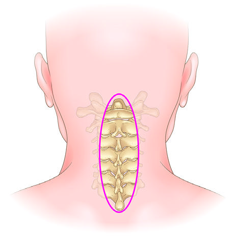
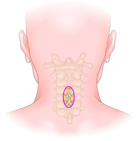
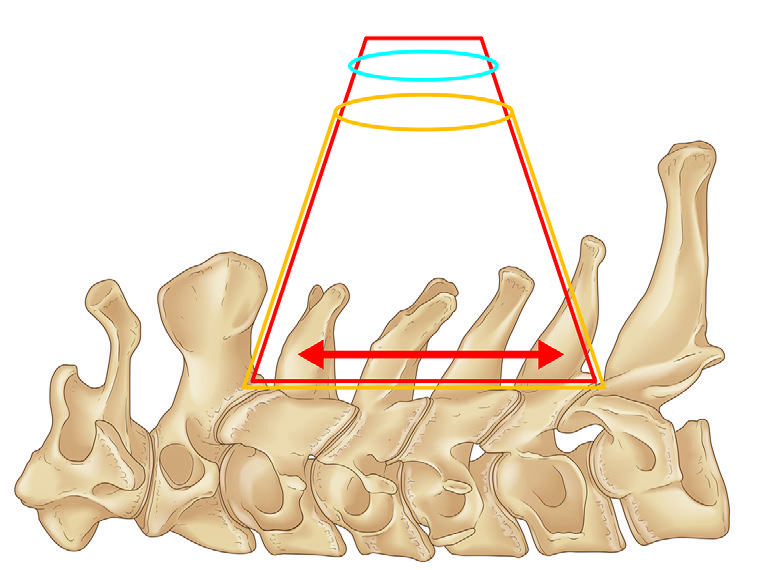
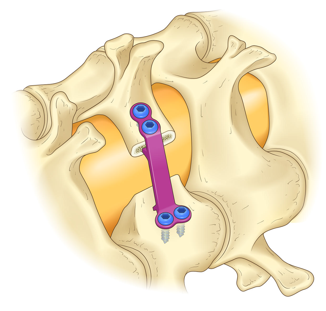
The figures below are cited from New OS NEXUS No.18. exLAP is treated as a related explanatory and implementation concept for OPERA/MIST-OPERA.
Original Technique Pages
The following pages are reproduced from pp.158-165 of the surgical technique chapter. To make the text and illustrations easier to read on screen, printing marks, page numbers, and lower print-production/footer information have been removed.
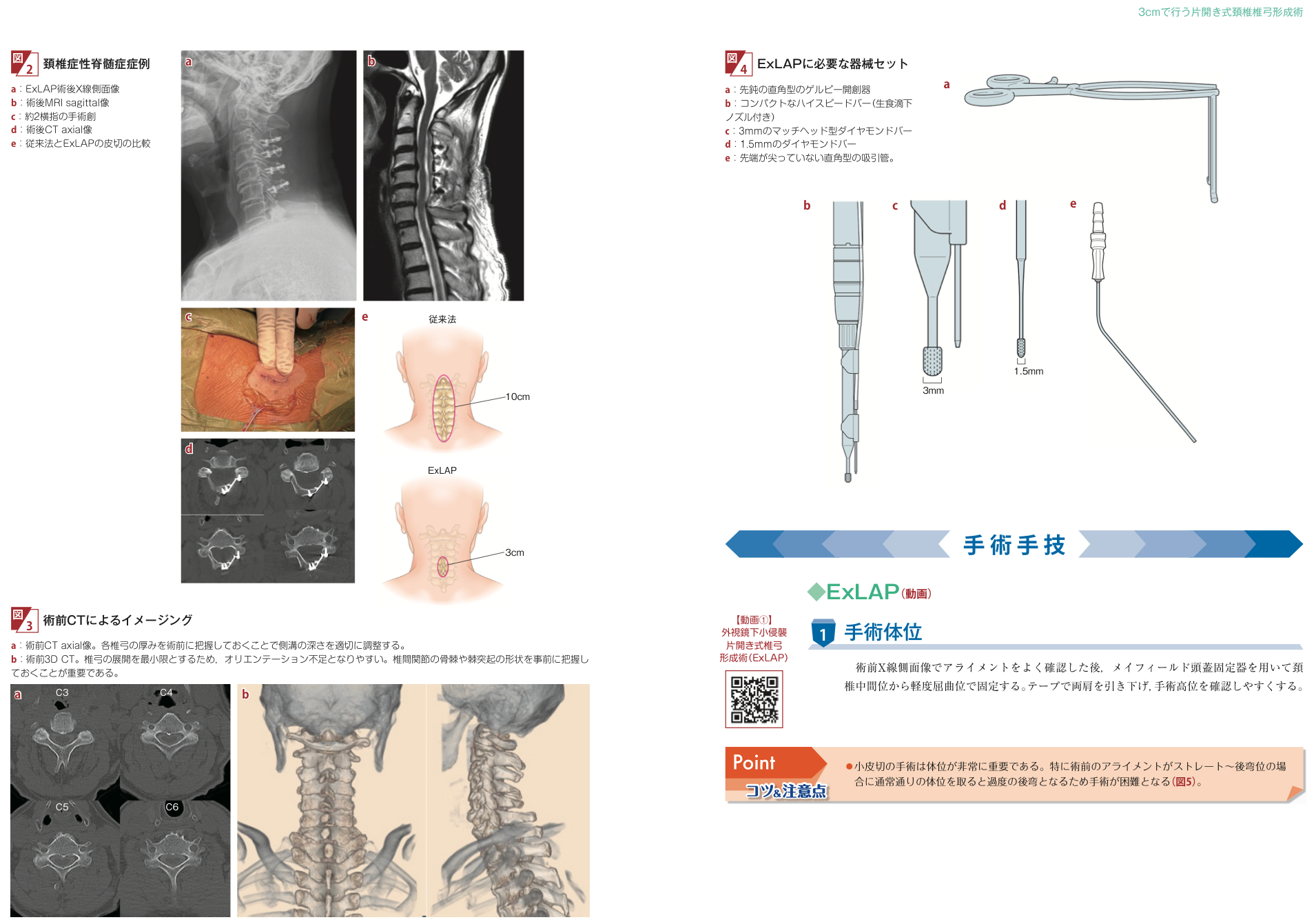
After confirming cervical alignment on a preoperative lateral X-ray, the head is fixed with a Mayfield clamp in a neutral to slightly flexed position. Both shoulders are taped downward so that the operative level can be checked more easily.
The technique uses a blunt right-angle Gelpi retractor, a compact high-speed burr with an irrigation nozzle, 3 mm and 1.5 mm diamond burrs, and a blunt right-angle suction tube. Preoperative axial CT is used to understand laminar thickness and adjust the depth of the lateral gutter. Three-dimensional CT is used to review spinous process shape and facet osteophytes before planning the working corridor.
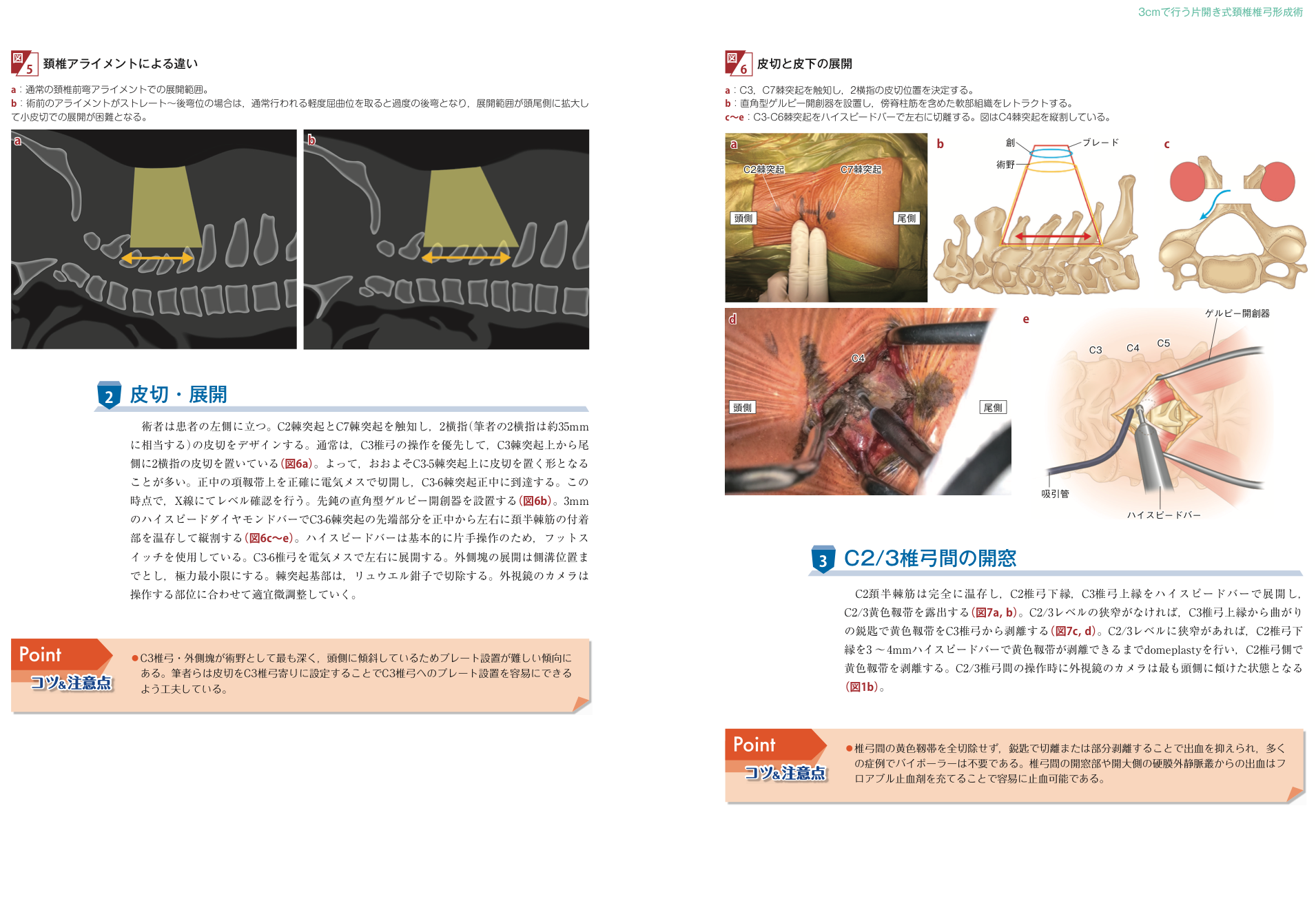
Positioning is especially important in a small-incision procedure. If the preoperative alignment is straight or kyphotic, routine slight flexion may create excessive kyphosis and make exposure through a small incision difficult.
The surgeon stands on the patient's left side. After palpating the C2 and C7 spinous processes, a two-fingerbreadth incision, about 35 mm for the authors, is designed. In many cases the incision is placed from the C3 spinous process caudally, roughly over C3 to C5. The midline nuchal ligament is opened precisely, the C3 to C6 midline is reached, and the level is confirmed by X-ray. A blunt right-angle Gelpi retractor is placed. The tips of the C3 to C6 spinous processes are split with a high-speed diamond burr while preserving the semispinalis cervicis attachments, and the laminae are exposed only as far laterally as needed for the lateral gutters.
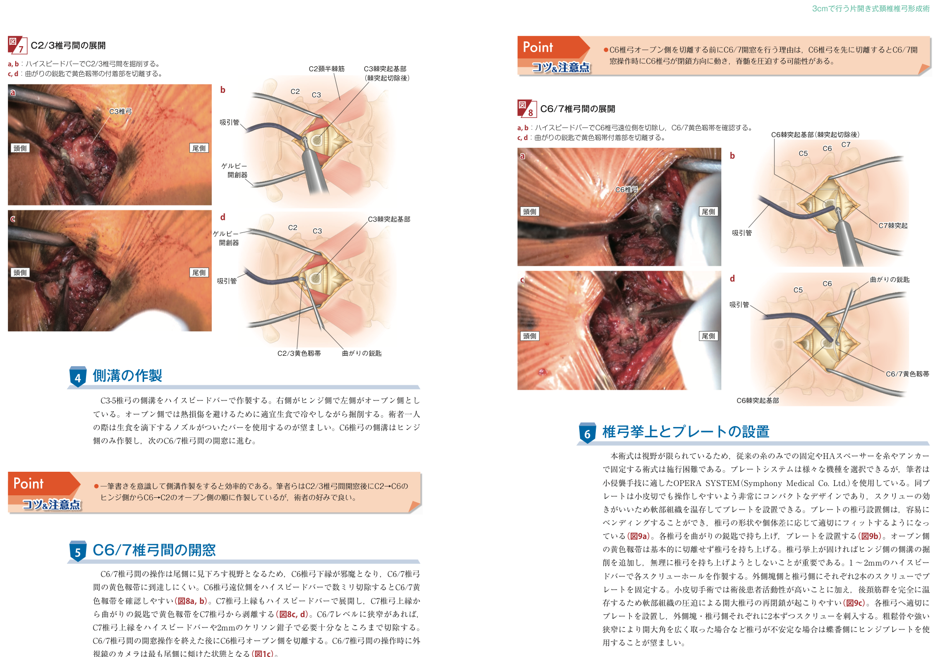
The C2 semispinalis cervicis is fully preserved. The lower edge of the C2 lamina and the upper edge of the C3 lamina are exposed with a high-speed burr to identify the C2/3 ligamentum flavum. If there is no C2/3 stenosis, the ligamentum flavum is detached from the C3 lamina with a curved curette. If C2/3 stenosis is present, a 3 to 4 mm domeplasty of the lower C2 lamina is performed until the ligament can be detached from the C2 side. During this step the exoscope camera is tilted most cranially.
The lateral gutters of the C3 to C5 laminae are made with a high-speed burr. The authors use the right side as the hinge side and the left side as the open side. On the open side, the bone is cooled intermittently with saline to avoid thermal injury; when operating alone, a burr with a saline-drip nozzle is preferred. For C6, only the hinge-side gutter is first created, and the procedure then moves to C6/7 fenestration. Because the C6/7 view looks caudally, the lower edge of C6 can obstruct access to the ligamentum flavum. Removing a few millimeters of the distal C6 lamina makes the C6/7 ligamentum flavum easier to identify. The upper edge of C7 is exposed, and the ligamentum flavum is detached from C7 with a curved curette. If C6/7 stenosis is present, the upper C7 lamina is removed sufficiently with a burr or 2 mm Kerrison rongeur. The open side of C6 is cut only after the C6/7 fenestration is complete.
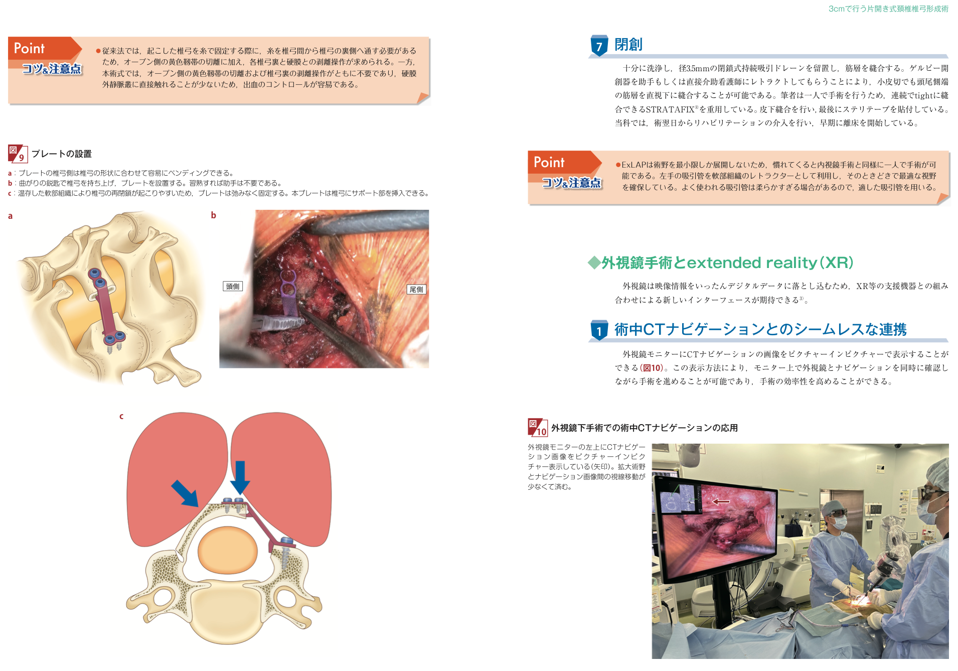
Because the field is limited, conventional suture-only fixation or hydroxyapatite spacer fixation can be difficult. The authors use the OPERA SYSTEM, which is compact and suited to minimally invasive manipulation. The laminar side of the plate can be bent to fit individual laminar shape. Each lamina is lifted with a curved curette and supported with a plate. The open-side ligamentum flavum is usually not cut. If elevation is stiff, additional drilling is performed on the hinge-side gutter; the lamina should not be forced upward. Each screw hole is prepared with a 1 to 2 mm burr, and the plate is fixed with two screws on the lateral mass side and two screws on the laminar side.
The laminar side of the plate can be bent to match laminar shape. After the lamina is raised with a curved curette, the plate is inserted and fixed without looseness because preserved soft tissue can promote reclosure. Unlike conventional suture fixation, this technique generally does not require passing sutures behind the lamina, cutting the open-side ligamentum flavum, or dissecting between the lamina and dura. Direct contact with the epidural venous plexus is reduced, making bleeding control easier.
After thorough irrigation, a 3.5 mm closed continuous suction drain is placed and the muscle layer is closed. Retraction of the Gelpi retractor by an assistant or scrub nurse allows the cranial and caudal ends of the muscle layer to be sutured under direct vision even through a small incision. Subcutaneous closure is followed by skin strips. Rehabilitation begins the day after surgery in the authors' department.
Because exoscope video is converted into digital data, integration with support devices such as XR is expected. CT navigation images can be displayed picture-in-picture on the exoscope monitor, allowing the surgeon to check the magnified surgical field and navigation image at the same time with less eye movement and improved operative efficiency.
Summary of the 28-patient exLAP study · 2024 original article · Surgical technique and figures
Positioning
For referrers, the key is not adding another name but making clear where OPERA sits within cervical laminoplasty.
Referral
Please include symptom course, neurological findings, comorbidities, medications, course of conservative treatment, MRI/CT/X-ray images, and prior surgical history. DICOM image sharing is preferred.
This page is general information for medical professionals. Individual indication is determined after examination, imaging, and review of general condition.