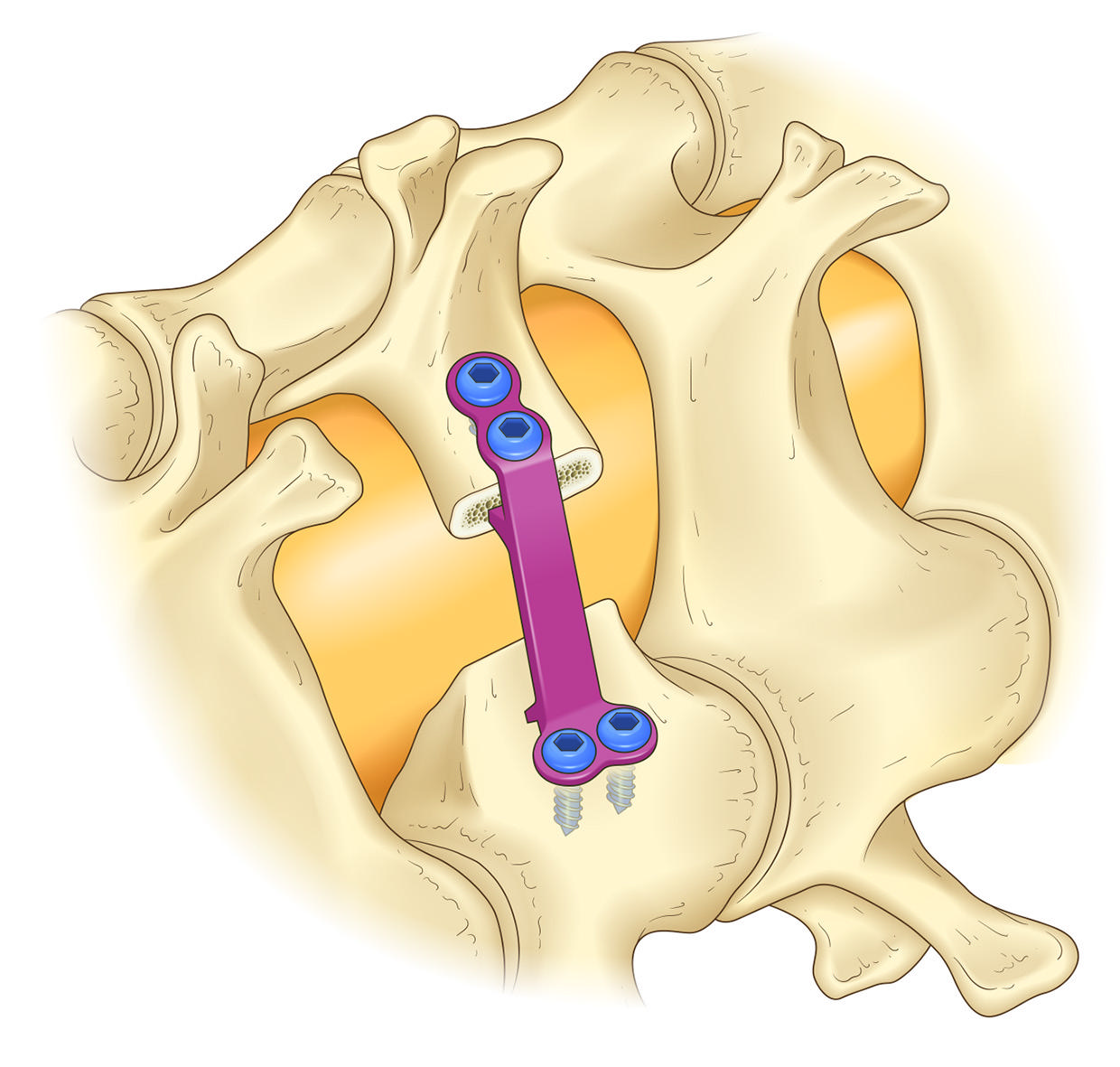
Textbook illustration of a plate supporting the opened lamina in open-door laminoplasty

OPEN-DOOR PLASTY FOR EARLY RECOVERY APPROACH

Cervical OPERA Method

Toward less invasive cervical surgery.

Understand the procedure. Explore the evidence. Consider your options with your doctor.

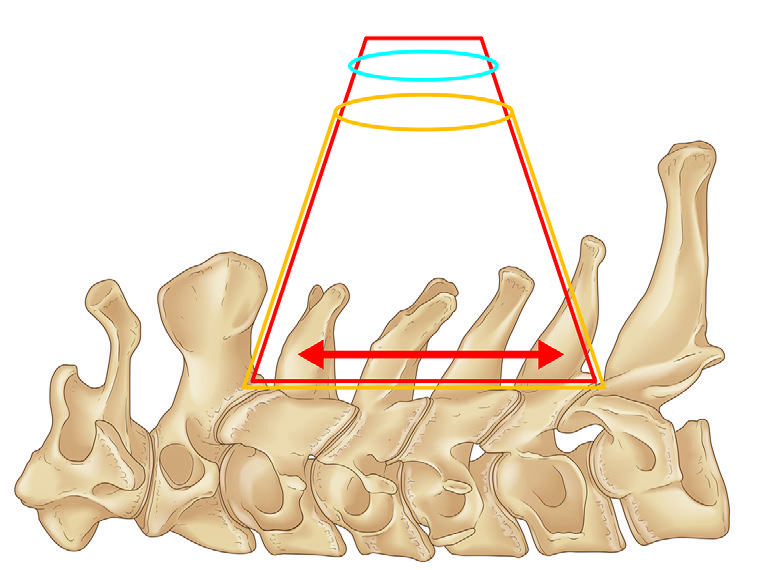
Illustration: OS NEXUS No.18, Fig.9. Reproduced with permission.

MIST-OPERA / exLAP

What is the OPERA method?

Information provided by: (Kameoka Municipal Hospital Spine Center)
Page updated:

Numbness in the hands, difficulty with fine movements, or trouble walking can sometimes come from compression of the spinal cord in the neck. This page walks through the Cervical OPERA Method by indication, differences from conventional surgery, risks, and possible consultation destinations.

The Cervical OPERA Method, also called MIST-OPERA, is a form of cervical laminoplasty considered for conditions such as cervical spondylotic myelopathy and ossification of the posterior longitudinal ligament. It combines a small incision, muscle-sparing exposure, microscope or exoscope visualization, and maintenance of the opened lamina in an open-door laminoplasty concept. Indication is judged by a physician based on neurological symptoms, imaging findings, and general condition. It is not suitable for every patient, and complications such as C5 palsy, pain, infection, bleeding, and reoperation must also be discussed.
01Check Symptoms

Numbness, hand clumsiness, and gait disturbance are reviewed.

02Review Images

MRI, CT, and X-rays show compression and bone shape.

03Compare Options

OPERA, conventional laminoplasty, anterior surgery, and fusion are compared.

04Find Instruction Links

Check hospitals where instructed physicians are affiliated.

Definition

First, clarify what kind of surgery it is

exLAP is the name used in the textbook and research papers for the exoscopic OPERA technique: exoscopic minimally invasive open-door laminoplasty. The Opera Laminoplasty System is the medical device used to maintain the position of the opened lamina, not the name of the operation.

Wataru Narita began developing the cervical OPERA method alongside the Opera Laminoplasty System in 2020 (information provided by the developer). The device approval number is 30500BZX00045000. The first package insert is dated April 1, 2023, and the third edition June 20, 2025. These are document publication and revision dates, not the approval date.

Main target diseases

  • Cervical spondylotic myelopathy
  • Ossification of the posterior longitudinal ligament (OPLL)
  • Multilevel cervical spinal cord compression

Actual indication depends on symptoms, MRI and CT findings, cervical alignment, and general condition.

Intended direction

The method aims to preserve the concept of broad cervical decompression while reducing surgical burden through a small incision, preservation of muscle attachments, magnified visualization, and laminar support.

When another approach may be better

Marked kyphosis, K-line negative OPLL, instability requiring fusion, infection, poor bone quality, or systemic risk may lead to consideration of another procedure.

Difference

How it differs from conventional cervical laminoplasty

The difference is not only the incision size. The concept links a small approach, visualization, soft-tissue preservation, and stable maintenance of the opened lamina.

Conventional open-door laminoplasty

  • Uses a wider exposure to secure the operative field
  • Visualization commonly relies on microscope or loupes
  • Fixation varies by method, including sutures, spacers, or plates
  • Requires explanation of axial pain, C5 palsy, infection, and other risks

OPERA / MIST-OPERA / exLAP

  • Designed to reach multiple laminae through a small incision
  • Uses microscope or exoscope visualization
  • Emphasizes reduced burden on muscle attachments and soft tissue
  • Maintains the opened lamina with a system such as the Opera Laminoplasty System

Textbook Figures

Key concepts shown with textbook figures

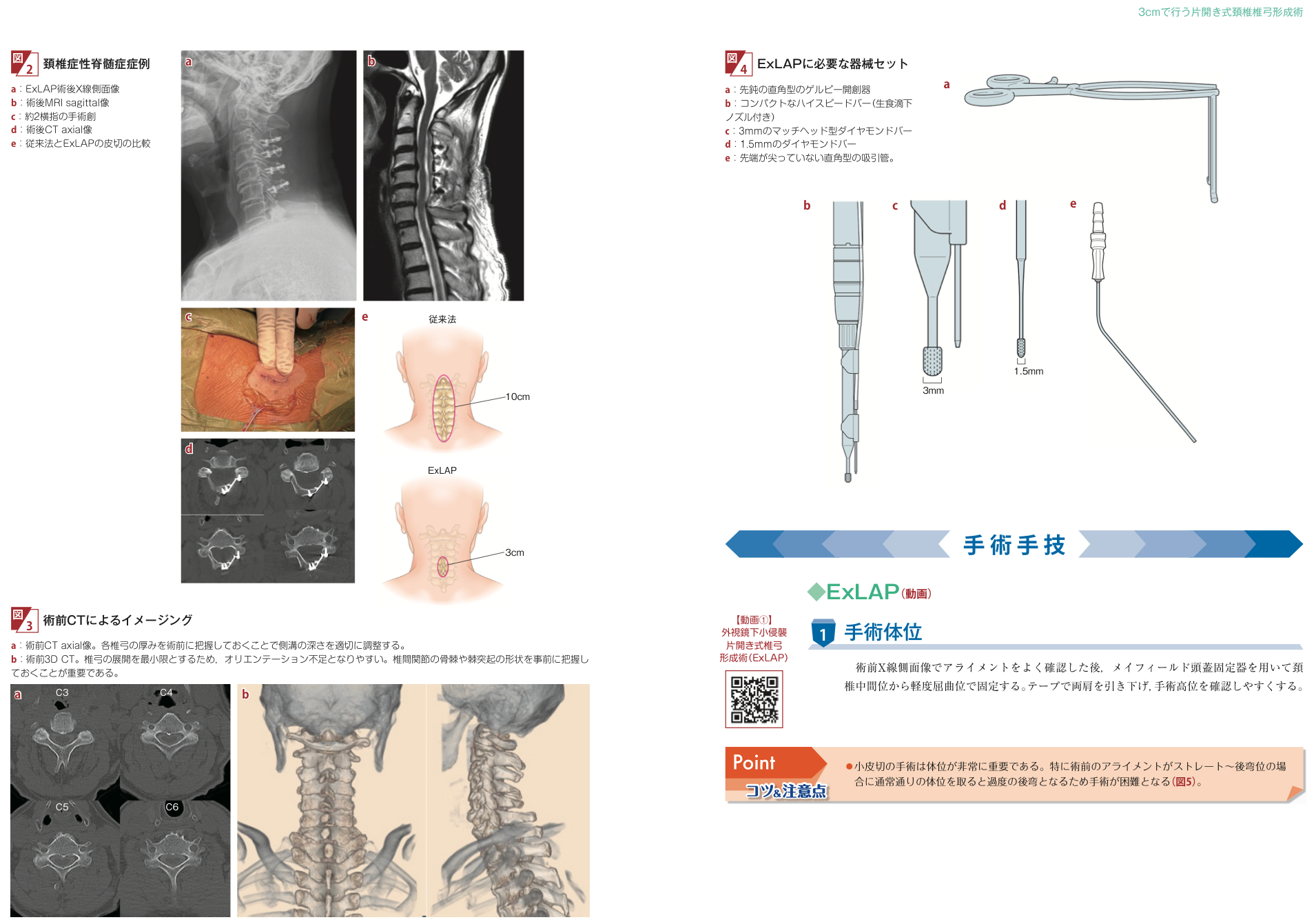
The following cited figures are from the chapter “Open-door cervical laminoplasty performed through a 3 cm incision: an attempt using exoscope and XR” in New OS NEXUS No.18.

Textbook figure showing the small-incision operative corridor
Operative corridor Preoperative CT helps assess laminar thickness and spinous process shape before planning the working corridor. Source: Yamane K, Narita W, New OS NEXUS No.18, Figure 3.
Textbook figure showing plate support for the opened lamina
Laminar support A concept image of maintaining the opened lamina with a plate. The device name is separated from the procedure name. Source: Figure 9.
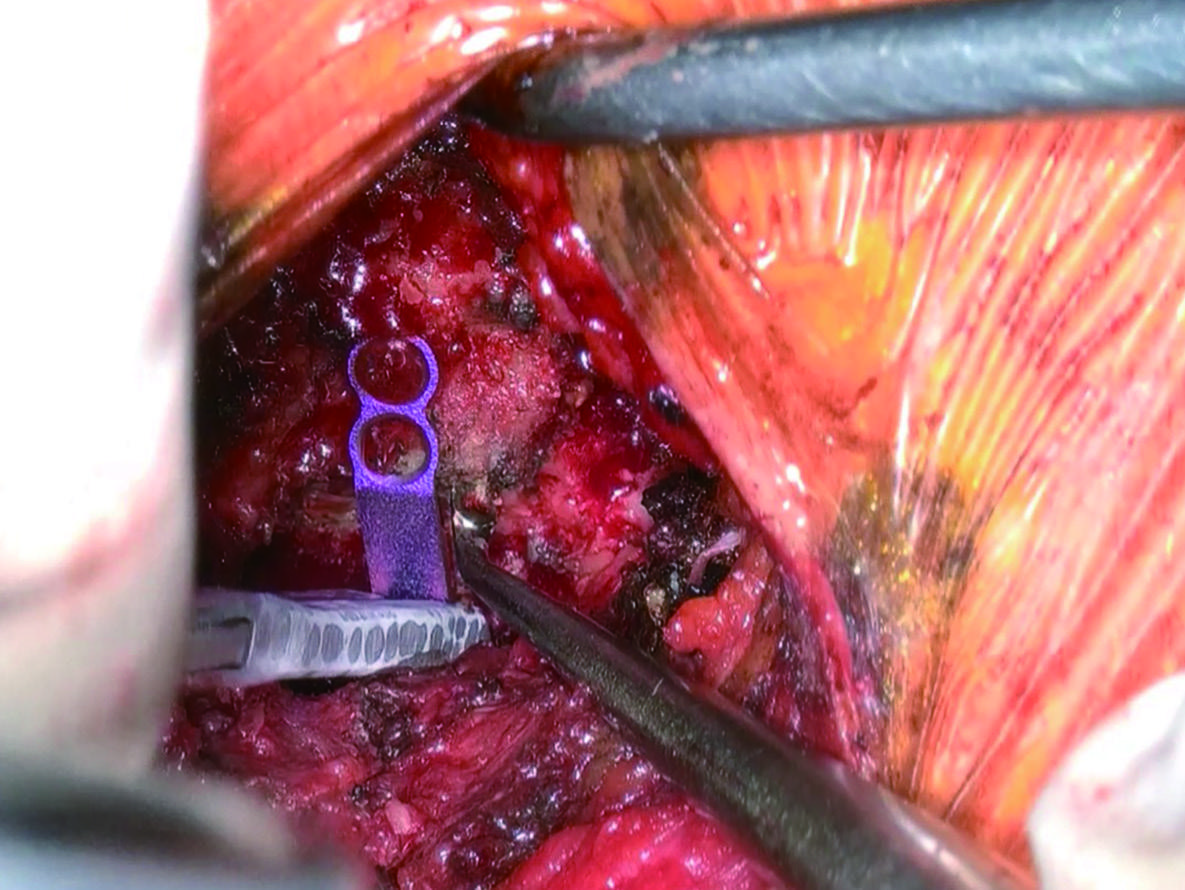
Textbook photograph showing plate placement
Plate placement Placement and screw fixation must be adapted to laminar anatomy. Source: Figure 9.
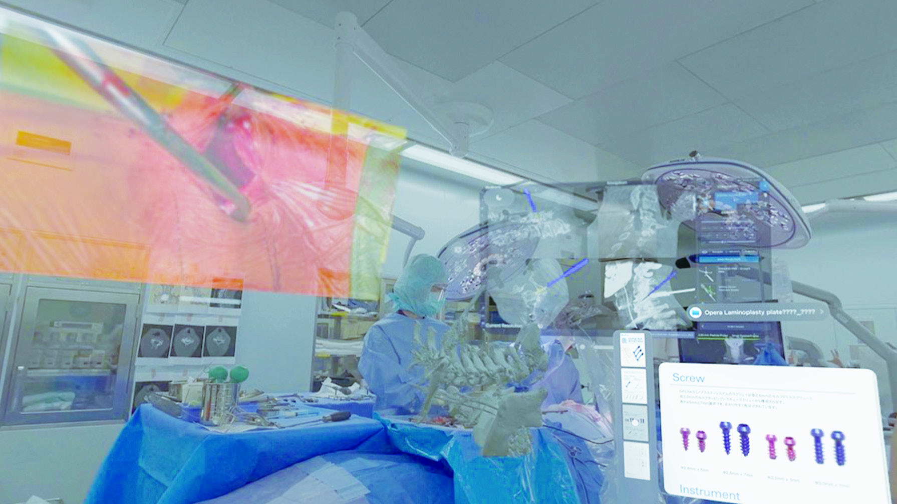
Textbook figure showing exoscope and XR integration
XR integration A future-facing concept for viewing exoscope images, navigation, and implant information in mixed reality. Device certification and latency remain important issues. Source: Figure 12.

Original Technique Pages

Surgical technique pages from OS NEXUS No.18

The following material is intended for physicians and healthcare professionals. Pages 158-165 of the surgical technique chapter are reproduced with printing marks, page numbers, and lower print-production/footer information removed so that the text and illustrations are easier to read.

Source: Kentaro Yamane and Wataru Narita, “Open-door cervical laminoplasty performed through a 3 cm incision: an attempt using exoscope and XR,” OS NEXUS No.18, pp.158-165, Medical View Co., Ltd. Reproduced with permission from Medical View Co., Ltd.; printing marks and related production information have been removed for readability.

pp.158-159

Surgical Position, Instruments, and CT-Based Preparation

Original source pages

Positioning

After confirming cervical alignment on a preoperative lateral X-ray, the head is fixed with a Mayfield clamp in a neutral to slightly flexed position. Both shoulders are taped downward so that the operative level can be checked more easily.

Instruments and imaging

The technique uses a blunt right-angle Gelpi retractor, a compact high-speed burr with an irrigation nozzle, 3 mm and 1.5 mm diamond burrs, and a blunt right-angle suction tube. Preoperative axial CT is used to understand laminar thickness and adjust the depth of the lateral gutter. Three-dimensional CT is used to review spinous process shape and facet osteophytes before planning the working corridor.

Technical note

Positioning is especially important in a small-incision procedure. If the preoperative alignment is straight or kyphotic, routine slight flexion may create excessive kyphosis and make exposure through a small incision difficult.

pp.160-161

Skin Incision, Exposure, and C2/3 Interlaminar Fenestration

Original source pages

Incision and exposure

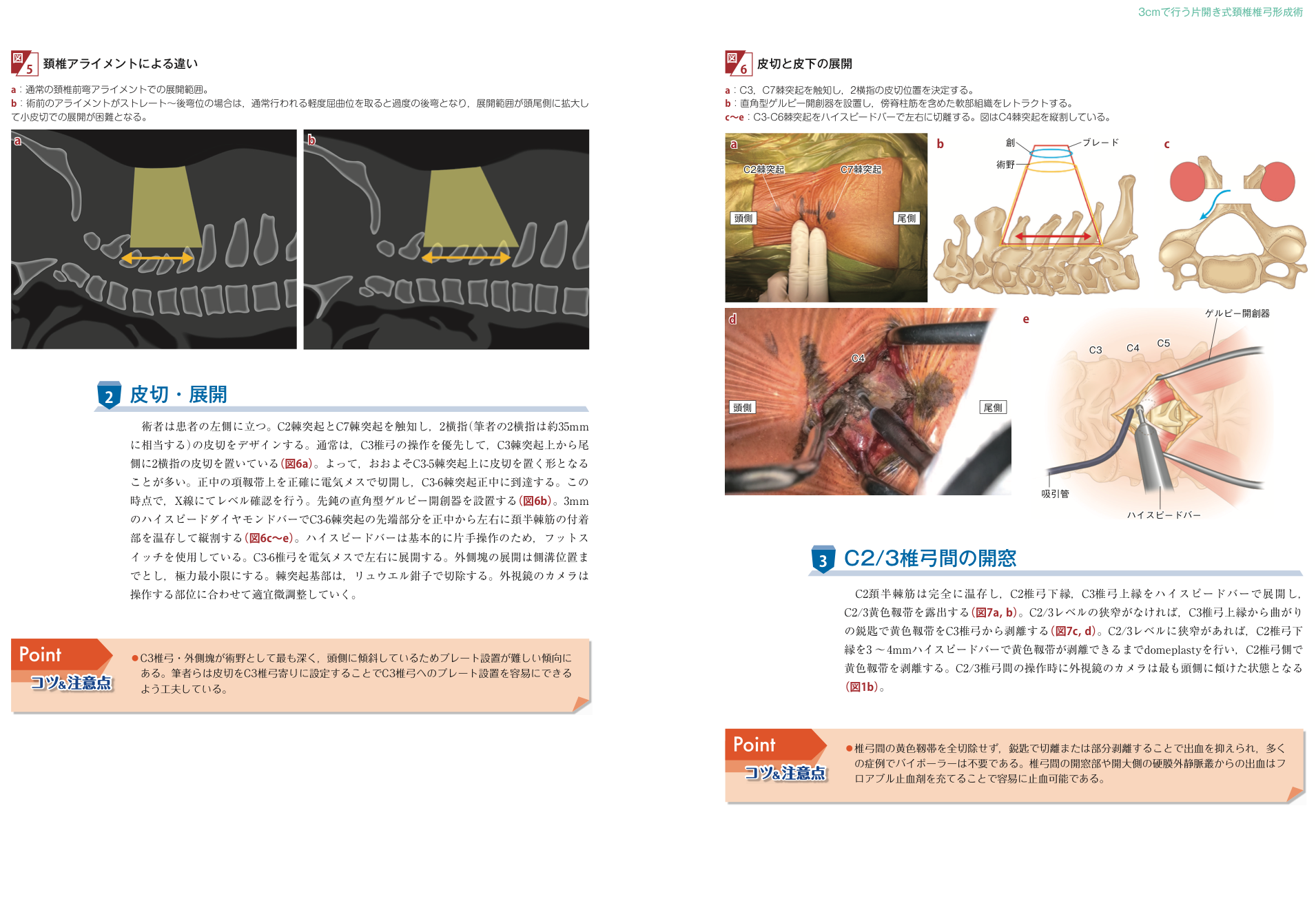
The surgeon stands on the patient's left side. After palpating the C2 and C7 spinous processes, a two-fingerbreadth incision, about 35 mm for the authors, is designed. In many cases the incision is placed from the C3 spinous process caudally, roughly over C3 to C5. The midline nuchal ligament is opened precisely, the C3 to C6 midline is reached, and the level is confirmed by X-ray. A blunt right-angle Gelpi retractor is placed. The tips of the C3 to C6 spinous processes are split with a high-speed diamond burr while preserving the semispinalis cervicis attachments, and the laminae are exposed only as far laterally as needed for the lateral gutters.

C2/3 fenestration

The C2 semispinalis cervicis is fully preserved. The lower edge of the C2 lamina and the upper edge of the C3 lamina are exposed with a high-speed burr to identify the C2/3 ligamentum flavum. If there is no C2/3 stenosis, the ligamentum flavum is detached from the C3 lamina with a curved curette. If C2/3 stenosis is present, a 3 to 4 mm domeplasty of the lower C2 lamina is performed until the ligament can be detached from the C2 side. During this step the exoscope camera is tilted most cranially.

Technical notes

  • The C3 lamina and lateral mass are deep and angled cranially, so plate placement tends to be difficult. Setting the incision closer to the C3 lamina can make C3 plate placement easier.
  • Instead of completely removing the interlaminar ligamentum flavum, cutting or partially detaching it with a curette can reduce bleeding. Bleeding from the fenestration or epidural venous plexus can usually be controlled with flowable hemostatic material.
pp.162-163

Lateral Gutters, C6/7 Fenestration, Decompression, and Plate Placement

Original source pages

Lateral gutters and C6/7 fenestration

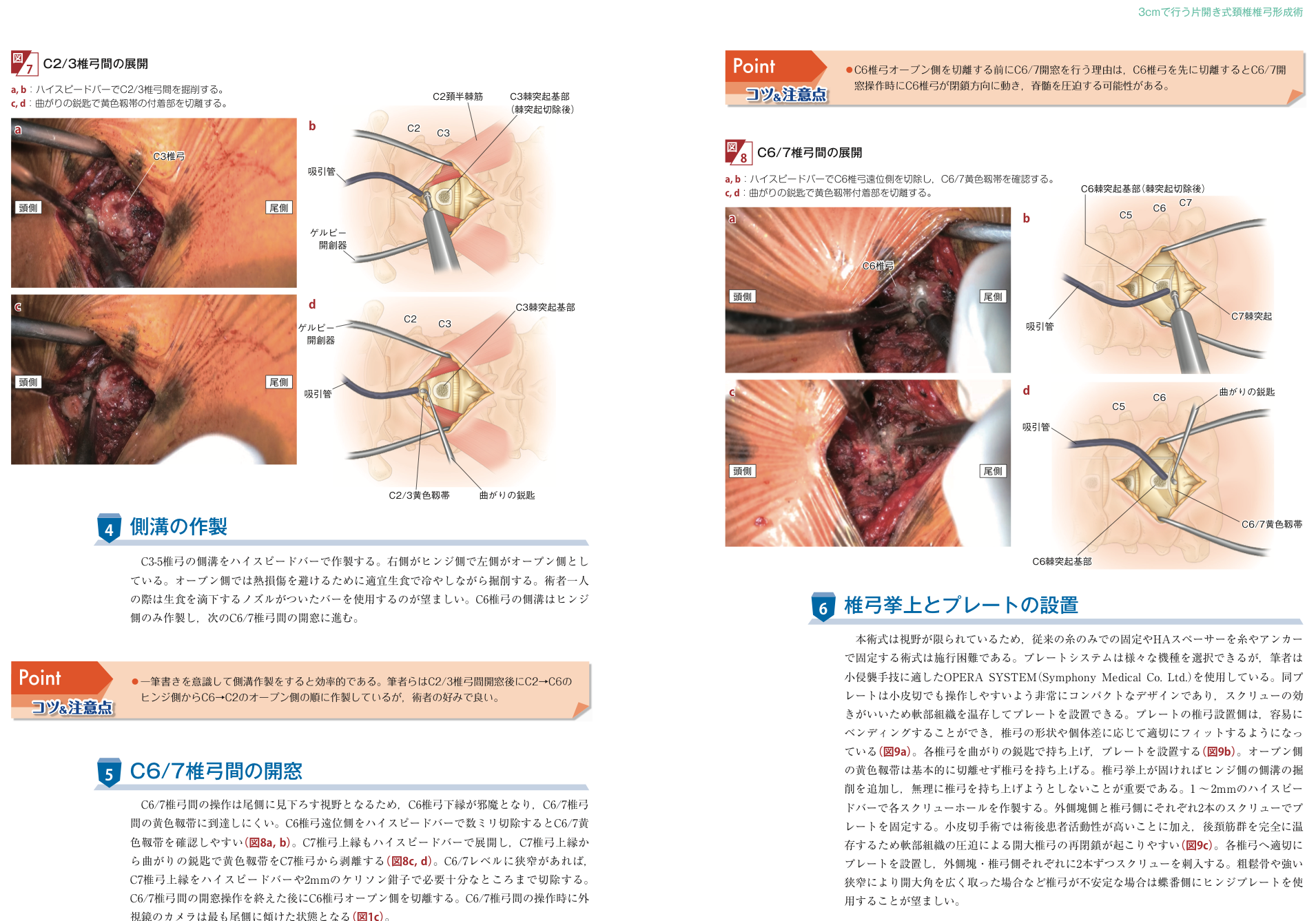
The lateral gutters of the C3 to C5 laminae are made with a high-speed burr. The authors use the right side as the hinge side and the left side as the open side. On the open side, the bone is cooled intermittently with saline to avoid thermal injury; when operating alone, a burr with a saline-drip nozzle is preferred. For C6, only the hinge-side gutter is first created, and the procedure then moves to C6/7 fenestration. Because the C6/7 view looks caudally, the lower edge of C6 can obstruct access to the ligamentum flavum. Removing a few millimeters of the distal C6 lamina makes the C6/7 ligamentum flavum easier to identify. The upper edge of C7 is exposed, and the ligamentum flavum is detached from C7 with a curved curette. If C6/7 stenosis is present, the upper C7 lamina is removed sufficiently with a burr or 2 mm Kerrison rongeur. The open side of C6 is cut only after the C6/7 fenestration is complete.

Plate-supported elevation

Because the field is limited, conventional suture-only fixation or hydroxyapatite spacer fixation can be difficult. The authors use the OPERA SYSTEM, which is compact and suited to minimally invasive manipulation. The laminar side of the plate can be bent to fit individual laminar shape. Each lamina is lifted with a curved curette and supported with a plate. The open-side ligamentum flavum is usually not cut. If elevation is stiff, additional drilling is performed on the hinge-side gutter; the lamina should not be forced upward. Each screw hole is prepared with a 1 to 2 mm burr, and the plate is fixed with two screws on the lateral mass side and two screws on the laminar side.

Technical notes

  • The authors create the gutters in a continuous motion, moving from the hinge side C2 to C6 and then from the open side C6 to C2 after C2/3 fenestration.
  • C6/7 fenestration is performed before cutting the open side of C6, because an early C6 cut may allow the lamina to move toward closure and compress the spinal cord.
  • With preserved posterior cervical muscles and higher postoperative activity, the opened lamina may tend to reclose. Stable plate fixation is therefore important; a hinge plate may be preferred when the lamina is unstable.
pp.164-165

Closure and Seamless Integration with Intraoperative CT Navigation

Original source pages

Plate fixation and closure

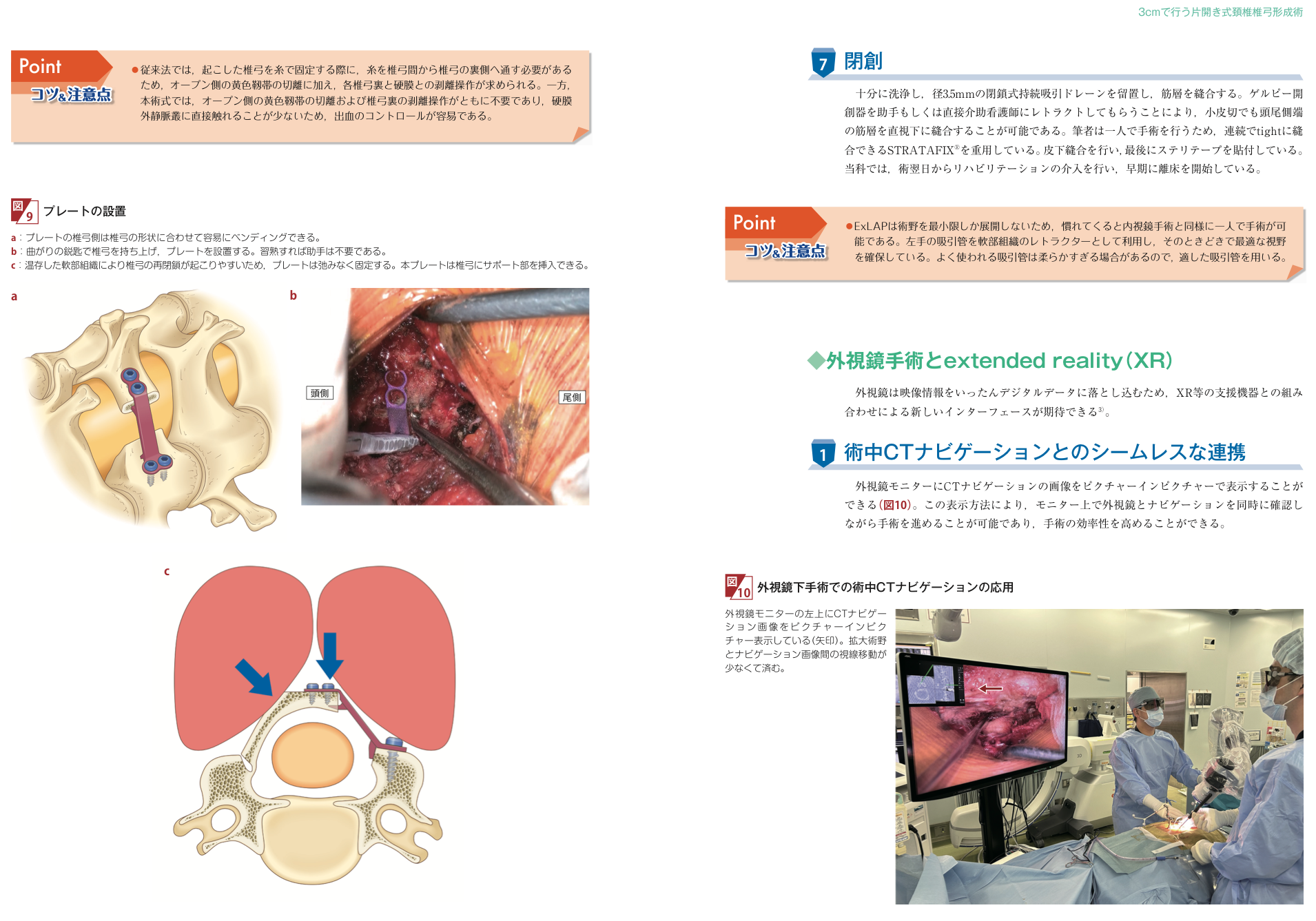
The laminar side of the plate can be bent to match laminar shape. After the lamina is raised with a curved curette, the plate is inserted and fixed without looseness because preserved soft tissue can promote reclosure. Unlike conventional suture fixation, this technique generally does not require passing sutures behind the lamina, cutting the open-side ligamentum flavum, or dissecting between the lamina and dura. Direct contact with the epidural venous plexus is reduced, making bleeding control easier.

After thorough irrigation, a 3.5 mm closed continuous suction drain is placed and the muscle layer is closed. Retraction of the Gelpi retractor by an assistant or scrub nurse allows the cranial and caudal ends of the muscle layer to be sutured under direct vision even through a small incision. Subcutaneous closure is followed by skin strips. Rehabilitation begins the day after surgery in the authors' department.

Intraoperative CT navigation and XR

Because exoscope video is converted into digital data, integration with support devices such as XR is expected. CT navigation images can be displayed picture-in-picture on the exoscope monitor, allowing the surgeon to check the magnified surgical field and navigation image at the same time with less eye movement and improved operative efficiency.

Patient Flow

From consultation to treatment decision

This page is not intended to rush treatment. It is an entry point for confirming symptoms and imaging, and for comparing surgery, conservative care, or another procedure.

Symptom review

Numbness, hand clumsiness, walking difficulty, falls, and bladder or bowel symptoms are reviewed.

Imaging review

MRI evaluates cord compression, CT evaluates ossification and laminar shape, and X-rays assess alignment or instability.

Procedure selection

The team compares OPERA with anterior surgery, fusion, conventional laminoplasty, or nonoperative care.

Risk discussion and consent

Expected benefits, complications, limitations, postoperative course, and possible reoperation are discussed.

Risks and limitations are described alongside advantages

  • C5 palsy, pain, residual numbness or weakness, infection, bleeding, dural injury, CSF leakage, implant-related issues, restenosis, and reoperation can occur.
  • A multicenter study of cervical laminoplasty reported severe C5 palsy in 43 of 1858 cases, or 2.3%.
  • A small-incision, early-recovery-oriented approach does not mean that every patient follows the same course.

Related Facilities

Hospitals where physicians instructed by Wataru Narita are affiliated

On this site, related facilities means hospitals where physicians who have received operative instruction from Wataru Narita are affiliated. It does not mean official certification, permanent availability of the OPERA method, or a designated referral destination. Please confirm appointment rules, referral requirements, clinic days, and physicians on each facility's official website.

Meaning

A guide for considering where consultation may be easier

For cervical disease, the appropriate treatment depends on symptoms, imaging, and general condition. This list is a practical cue for patients comparing consultation destinations by region and access.

Wataru Narita operative instruction Physicians at each hospital confirm local system Possible consultation destination
TohokuSendai Nishitaga Hospital

Sendai, Miyagi

KantoSangubashi Spine Surgery Hospital

Shibuya, Tokyo

KantoTokyo Spine and Joint Clinic Haneda

Ota, Tokyo

ChubuMishima Central Hospital

Mishima, Shizuoka

ChugokuOkayama Medical Center

Okayama City

ShikokuKochi Health Sciences Center

Kochi City

Chugoku / Okayama

National Hospital Organization Okayama Medical Center

Located in Kita-ku, Okayama. Access from the Okayama Station area is mainly by bus or car; the official page provides bus, road, and parking information.

View access

Tohoku / Miyagi

National Hospital Organization Sendai Nishitaga Hospital

Located in Taihaku-ku, Sendai. From Sendai Station, take a Miyagi Kotsu bus for about 30 minutes and walk about 5 minutes from Sendai Nishitaga Hospital-mae.

View access

Shikoku / Kochi

Kochi Health Sciences Center

Located in Kochi City. About 15 minutes by taxi or about 30 minutes by bus from JR Kochi Station. The hospital states that outpatient visits are generally by appointment.

View access

Kanto / Tokyo

Shunyo-kai Sangubashi Spine Surgery Hospital

Located in Shibuya, Tokyo. About 5 minutes on foot from Sangubashi Station on the Odakyu Line, or 8 minutes from JR Yoyogi Station.

View hospital information

Kanto / Tokyo

Tokyo Spine and Joint Clinic Haneda

2-20-25 Higashi-Rokugo, Ota-ku, Tokyo. About 6 minutes on foot from Zoshiki Station on the Keikyu Main Line; bus access from the JR Kamata Station area is also listed.

View access

Chubu / Shizuoka

Shijinkai Mishima Central Hospital

Located in Mishima, Shizuoka. About 5 minutes by car from the south exit of Mishima Station, or 2 minutes on foot from Mishima-Hirokoji Station.

View overview and access

Sources

Main sources

Kameoka Municipal Hospital Spine Center

The public hospital page describes MIST-OPERA as Open-door Plasty for Early Recovery Approach and lists Dr. Wataru Narita and the Spine Center.

Kameoka Municipal Hospital

Yamane K, Narita W, et al. J Clin Med. 2024

A technical note and preliminary clinical study of exLAP in 28 cases, including acute postoperative neck pain.

doi:10.3390/jcm13082173

Opera Laminoplasty System

OPENOVA and the package insert identify the implant, manufacturer, generic device name, and approval number 30500BZX00045000.

OPENOVA Online

Frequently asked questions about the OPERA method

What is the cervical OPERA method?

The cervical OPERA method (MIST-OPERA) is an open-door cervical laminoplasty that emphasizes a small incision and preservation of muscle attachments. It widens the space for the spinal cord from the back of the neck. The exoscopic technique is called exLAP.

Which conditions may be suitable?

It may be considered for cervical spondylotic myelopathy or ossification of the posterior longitudinal ligament (OPLL). Symptoms, the compression level, spinal alignment, instability and general health guide the choice between nonsurgical treatment and different operations.

Does a small incision eliminate complications?

No. Possible complications include C5 palsy, neck pain, infection, bleeding, neurological injury and reoperation. A small incision does not eliminate these risks.

When can I leave hospital or return to work?

There is no single timetable. Neurological symptoms, general health, postoperative recovery and the demands of your work determine the plan with your treating physician.

What research is available?

A preliminary 2024 study compared acute postoperative outcomes in 17 exLAP patients and 11 conventional laminoplasty patients, 28 in total. It does not establish long-term results or effectiveness for every patient.

Do all related hospitals offer OPERA surgery?

Related facilities are hospitals where physicians instructed by Wataru Narita work. Confirm current availability, the treating physician, referral requirements and appointments with each hospital.

Who can explain the cost and insurance coverage?

Ask the hospital where you plan to receive treatment. The procedure, admission length, insurance eligibility and cost-sharing affect the amount payable. An implant price is not the total cost of surgery and hospitalization.

Consultation

Consultation and referral

Whether the Cervical OPERA Method is appropriate is determined by symptoms and imaging. A referral letter, MRI, CT, and X-ray images are helpful for evaluation.

Last updated: October 2, 2026. This page is general medical information and does not replace individual diagnosis.